Science
AI Revolutionizes 3D Reconstruction of Biological Structures
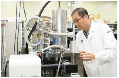
The Korea Research Institute of Standards and Science (KRISS) has introduced an innovative artificial intelligence (AI) image segmentation algorithm that can efficiently reconstruct three-dimensional (3D) structures from two-dimensional (2D) cross-sectional images. This advancement utilizes data captured by scanning electron microscopes (SEM), marking a significant leap forward in the field of microscopy and biological imaging.
The algorithm developed by KRISS allows researchers to quickly generate detailed 3D models from the often complex and intricate 2D images produced by electron microscopy. This technology not only enhances the speed of reconstruction but also improves the accuracy of the models, enabling scientists to gain deeper insights into the microstructural properties of biological samples.
Impact on Research and Industry
According to Lee Ho Seong, the president of KRISS, the new algorithm has the potential to transform various research fields, including materials science and biology. By streamlining the process of visualizing microscopic structures, researchers can focus more on analysis and interpretation rather than spending excessive time on image processing. This efficiency could lead to faster discoveries in areas such as drug development and disease understanding.
The ability to reconstruct 3D structures from 2D images also opens up new avenues for industries reliant on high-resolution imaging. These include pharmaceuticals, biotechnology, and nanotechnology sectors, where precise visualization of materials at a microscopic level is critical for product development and quality control.
Technological Details and Future Applications
The AI algorithm is designed to learn from large datasets, improving its performance over time. This machine learning approach allows it to adapt to various types of biological samples and imaging conditions. As a result, the algorithm can handle diverse challenges presented by different specimens, ensuring robust performance across applications.
In practical terms, this means that researchers can analyze complex biological phenomena—such as cellular interactions, tissue architecture, and pathogen structures—more effectively than ever before. The implications for healthcare, particularly in understanding diseases at a cellular level, could be profound, paving the way for innovative treatment strategies.
As KRISS continues to refine this technology, collaboration with academic institutions and industry partners is expected to accelerate its deployment in real-world applications. The institute aims to share its findings and technology to foster further advancements in microscopy and related fields.
The development of this AI-based reconstruction algorithm exemplifies how cutting-edge technology can enhance scientific research, bridging the gap between complex data and actionable insights. As the field of microscopy evolves, tools like these will play a crucial role in advancing our understanding of the microscopic world.

Urgent Warning: Middle Managers Face Job Cuts Amid Tech Shakeup

Innovative Leaf-Scanning Technology Helps Farmers Assess Fruit Ripeness

Houston Cougars Face Arkansas Razorbacks in High-Stakes Showdown

Incyte’s Antibody Shows Promise for Treating Rare Blood Cancer

Parents Face Financial Strain by Supporting Adult Children

Democrats Capitalize on “Affordability” to Shift Political Landscape

Aimee Ng Revitalizes the Frick Collection with Curatorial Innovation

Master the Art of Folding Fitted Sheets: A Simple Guide

Russia Accelerates INSTC to Bypass Sanctions and Boost Trade

Inventor Achieves Breakthrough with 2 Billion FPS Laser Video

Community Unites for 7th Annual Into the Light Walk for Mental Health

Charlie Sheen’s New Romance: ‘Glowing’ with Younger Partner

Dua Lipa Aces GCSE Spanish, Sparks Super Bowl Buzz with Fans

Curium Group, PeptiDream, and PDRadiopharma Launch Key Cancer Trial

Mother Fights to Reunite with Children After Kidnapping in New Drama

Former Mozilla CMO Launches AI-Driven Cannabis Cocktail Brand Fast
R&B Icon D’Angelo Dies at 51, Leaving Lasting Legacy

Israel Reopens Rafah Crossing After Hostage Remains Returned
-
 Science2 months ago
Inventor Achieves Breakthrough with 2 Billion FPS Laser Video
-
 Health2 months ago
Community Unites for 7th Annual Into the Light Walk for Mental Health
-
 Top Stories2 months ago
Charlie Sheen’s New Romance: ‘Glowing’ with Younger Partner
-
 Entertainment2 months ago
Dua Lipa Aces GCSE Spanish, Sparks Super Bowl Buzz with Fans
-
 Health2 months ago
Curium Group, PeptiDream, and PDRadiopharma Launch Key Cancer Trial
-
 Entertainment2 months ago
Mother Fights to Reunite with Children After Kidnapping in New Drama
-
 Top Stories2 months ago
Former Mozilla CMO Launches AI-Driven Cannabis Cocktail Brand Fast
-

 World2 months ago
R&B Icon D’Angelo Dies at 51, Leaving Lasting Legacy
-
 World2 months ago
Israel Reopens Rafah Crossing After Hostage Remains Returned
-

 Health2 months ago
YouTube Launches New Mental Health Tools for Teen Users
-

 Business2 months ago
Tyler Technologies Set to Reveal Q3 Earnings on October 22
-

 Entertainment2 months ago
Red Sox’s Bregman to Become Free Agent; Tigers Commit to Skubal